Aflatoxins were initially isolated and identified as the causative toxins in Turkey X disease (necrosis of the liver) in 1960 when over 100,000 turkeys died in England (Asao, 1963). They are the most studied mycotoxins with over 5,000 research papers published through 2002. There are four generally recognized Aflatoxins designated B1, B2, G1 and G2 (Figures 1 and 2).
The metabolites, M1 & M2, are found in milk (Thirumala-Devi et al (2002).
The order of toxicity is B1 greater than G1, greater than G2, greater than B2. (IARC, 1976). However, Aflatoxin B1 is the major mycotoxin produced by most species under culture conditions (Ciegler & Bennet, 1980). Because of this and its toxicity, B1 is the most frequently studied of the four.
Aflatoxins are produced by different species of Aspergillus, particularly flavus, oryzae, fumigatus and parasiticus, as well as members of the genera Penicillium (El-Naghy et al, 1991; Searle 1976; Aflatoxins 2002). Strains of Aspergillus flavus and parasiticus produce mycotoxins under favorable conditions.
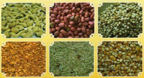
Aflatoxins can contaminate corn, cereals, sorghum, peanuts and other oil-seed crops. Thus, food contamination by this group of mycotoxins has been implicated in both animal and human Aflatoxicosis.
In addition, inhalation of aflatoxins is associated with disease and injury in both animals and humans. Finally, aflatoxins are known animal carcinogens and have been linked to cancer of the liver in humans and kidneys in rodents (Markow et al, 1973; Epstein et al, 1969; Sun et al, 2001; Wild & Turner, 2002).
Aflatoxins and Cancer in Humans
Aflatoxins are carcinogenic to humans and animals. Overall summary evaluation of carcinogenic risk to humans is Group 1 (IARC, 1976; Searle, 1976; Dominguez-Malagon & Gaytan-Graham (2001).
Aflatoxin B1 is a potent liver carcinogen in a variety of experimental animals. It causes liver tumors in mice, rats, fish, marmosets, tree shrews and monkeys following administration by various routes. Types of cancers described in research animals include hepatocellular carcinoma (rats), colon and kidney (rats), cholangiocellular cancer (hamsters), lung adenomas (mice), and osteogenic sarcoma, adenocarcinoma of the gall bladder and carcinoma of the pancreas (monkeys) (IARC, 1976).
In humans, Aflatoxin B1 has been linked to hepatocellular carcinoma from three studies reported in the medical literature, as follows:
An increased incidence (10 % excess) of hepatocellular carcinoma was reported in the southeastern portion of the U.S. in areas of high daily intake of Aflatoxin B1. The daily intake of B1 in the southeastern subjects was 13-197 ug/kg body weight as compared to those in northern and western areas with a daily intake of 0.2-0.3 ug/kg body weight (IARC, 1987).
In China, a strong correlation between the intake of peanut, peanut oil and corn and increased mortality rates for liver cancer were reported in five groups of inhabitants from four villages. The mortality rates were 125, 97.40, 41.65, 24.01 and 1.05, respectively. The median intake of aflatoxin B1 for each group was 6.05, 6.36, 2.69, 1.83 and 0 ug/day. The median daily urine concentrations of M1 metabolite were 16.46, 8, 29, 4.78 and 1.21 ng/person. A significant correlation was found between the mortality rates of primary liver cancer and intake of aflatoxin B1. Further, analysis of M1 in the urine can be used as an index for human exposure to aflatoxin B1 in an epidemiological study (Aflatoxins, 2002).
Cancer in 67 men who had inhaled particles contaminated with aflatoxin were reported in an 11-year follow up study. They worked in a mill crushing peanuts and other oil seeds. Two of the men developed fatal liver disease, while eleven developed cancers of various organs. The 13 men had inhaled doses estimated to be between 160 to 395 ug/cubic meter/man/wk. In 55 matched control men, 4 developed cancer and none died from liver disease. The excess cancers in this study was not significant, however the number of subjects was insufficient to exclude a significant positive correlation (Aflatoxins, 2002; IARC,1976).
Finally, it was reported that in hepatocellular carcinoma cases exposed to aflatoxin B1, mutation of p53 gene is fixed at codon 249 third base and takes the form of G to T transversion. It appears from the reported observations that it is a definite marker of mutation, which is induced by aflatoxin B1 mutagen and is applicable for molecular epidemiology survey of sufferers of aflatoxin B1 exposure among hepatocellular carcinoma cases (Deng & Ma, 1998).
Aflatoxicosis in Humans
Suspected cases of Aflatoxicosis have been reported in India. Over 200 villages in western India experienced an outbreak of disease affecting humans and dogs. The illness was characterized by jaundice, rapidly developing ascites, portal hypertension and a high mortality rate. Death usually occurred from massive gastrointestinal bleeding. The disease was confined to the very poor who ate badly molded corn containing aflatoxin at a concentration of 6.25 to 15.6 ppm. The average daily intake was 1-6 mg of aflatoxin (Aflatoxins, 2002; Keeler et al, 1983).
Reye's syndrome is characterized by symptoms of encephalopathy and fatty degeneration of the viscera. It occurs in children under 16 and is believed to follow nonspecific viral illness, influenza B and varicella. Abnormalities are present in mitochondrial structure and function, including defective oxidative phosphorylation (Stein, 1990).
Aflatoxins have been reported to be associated with a Reye's-like Syndrome in Thailand, New Zealand, Czechoslovakia, the United States (Aflatoxins, 2002; Ryan et al (1979) Malaysia (Chao, 1991), Venezuela (Burggera, 1986) and Europe (Dvorackova et al, 1977, Stora et al, 1983).
An incident of 106 fatal cases of hepatic disease among 397 individuals who became ill after eating maize contaminated with aflatoxin also suggests that acute aflatoxicosis can occur in human beings (Haddad, 1990). Recently, outbreaks of aflatoxicosis resulting from contaminated grain occurred in Kenya (Azziz-Baumgartner et al, 2005; Lewis et al, 2005).
Post-weaning exposure to aflatoxin-contaminated maize has resulted in impaired growth of African children consuming the grain vs infants still being breast fed.
Aflatoxin concentration mean values were 40.4, 10.1, 8.7 pico grams/mg in maternal, cord and infant blood. In utero exposure also resulted in growth faltering in Gambian infants (Gong et al, 2004; Turner et al, 2007).
The Reye's-like syndrome reported in various places around the world was characterized by multiple symptoms and clinical findings that included disturbed consciousness, fever, convulsions, vomiting, disturbed respiratory rhythm, altered muscle tone and altered reflexes. Serum glutamic-pyruvic transaminase and glutamic-oxalacetic acid transaminase (mitochondrial) enzymes levels were elevated. Hypoglycemia and low cerebrospinal fluid glucose were observed. The onset of the illness included coughing, rhinorrhea, sore throat, earache, slightly enlarged, firm yellow liver, and a pale, slightly widened renal cortex. A high rate of mortality occurred in 81 % of the diagnosed cases.
Since Reye's Syndrome is characterized by abnormal mitochondrial structure and function, it is of interest to note that aflatoxin B1 causes abnormal mitochondrial structure and function (Shanks et al, 1986; Rainbow et al, 1994; Obasi. 2001; Pasupathy et al, 1999; Sajan et al, 1996).
Aflatoxins have been demonstrated in human cord blood and sera of women immediately after birth. These results demonstrated the transplacental transfer and concentration of aflatoxin by the fetal-placental unit (Aflatoxins, 2002; Turner et al, 2007). Moreover, neonates with and without jaundice were investigated for the presence of aflatoxins in the cord blood. Jaundice and decreased birth weight were correlated with a higher concentration of aflatoxin B1. The observations showed that neonates are exposed to aflatoxin prenatally and that high incidence of jaundice occurred in wet warm months (Abulu et al, 1998). These observations are of biological significance for humans because aflatoxins are mutagenic, carcinogenic, teratogenic and immunosuppressive in animals. The implications of these findings are potentially profound and deserve further study (Aflatoxins, 2002)
Three cases of pulmonary interstitial fibrosis, two agricultural workers and one textile worker, with exposure to aflatoxins have been reported (Dvorackova & Pichova, 1986). Lung samples from the three workers contain aflatoxin B1 suggesting a possible occupational exposure of aflatoxin via the respiratory tract and lung disease.
Immune Response in Humans and Aflatoxins
Antibodies to aflatoxin B1 have been reported in humans. At present, these antibodies are considered indicative of exposure and may or may not be related to disease (Ryan et al, 1979; Wang et al, 2001).
In addition, genetic polymorphism in humans lacking glutathione transferase enzymes, cytochrome CYP3A5 and microsomal epoxide hyrdolase (mEH) enzymes along with dietary intake are associated with increased levels of aflatoxin-albumin adducts. These observations are supportive of the production of antibodies to aflatoxin-albumin adducts.
When detoxification pathways either lack (null condition) or have mutations in certain enzymes, free radicals increase leading to protein-free radical adducts Chen et al, 2000; Wild et al, 2000; Wojnowski et al, 2004; Dash et al, 2007).
Workers handling animal feed exposed to mycotoxins, including aflatoxins, were examined for alterations in plasma alpha tumor necrosing factor (TNF-alpha). Airborne concentration of aflatoxin was 0.99 ng/m3. Exposed workers had increased levels of TNF-alpha and concomitant changes in serum lactate dehydrogenase isoenzyme activity (Nuntharatanpong et al, 2002).
Aflatoxicosis in Animals
Domestic animals (pets and agricultural), monkeys, laboratory rats and mice have been the subject of a large body of research on the adverse effects of aflatoxins (particularly B1). These effects include adducts and mutations, cancer, immunosuppression, lung injury and birth defects. Also, aflatoxins have been shown to interact with DNA (nuclear and mitochondrial adducts) and polymerases responsible for DNA and RNA synthesis. The literature is voluminous and can be researched through the National Library of Medicine. Thus, aflatoxicosis in animals will be briefly reviewed here.
Aflatoxins are immunosuppressive in a variety of animals making them more susceptible to infection by various microorganisms. The animals include sheep, cattle, mice, rats rabbits, pigs and poultry, among others (Aflatoxins, 2002; Sharma, 1992; Silvott, et al,1997; Dimitri & Gabal, 2996; Jakab et al, 1994).
Aflatoxicosis in horses, cows and dogs has been described. Essentially, the disease process is similar to that seen in humans. However, because of the ability to study animals in more detail in the laboratory, considerable more scientific information has accumulated regarding the course of the disease.
Calves develop a disease that features blindness, circling and falling down, twitching of ears and grinding of teeth. Spasm of the rectum is seen in most cases. Death usually follows within two days of onset of severe clinical signs. Postmortem findings revealed pale, firm and fibrosed liver. Histologically the main liver changes are centrilobular necrosing, bile duct proliferation and veno-occlusive disease. The kidneys are yellow and surrounded by wet fat. Ascites and edema of the mesentery (enteritis) and rectal eversion are common findings.
Other pathological features in cattle are blood coagulation defects which may involve impairment of prothrombin, factors VII and X and possibly factor IX.
A single dose of aflatoxin causes increases in plasma enzymes (aspartate aminotransferase, lactate dehydrogenase, glutamate dehydrogenase, gamma-glutamyltransferase and alkaline phosphatase and in bilirubin, probably reflecting liver damage. Other abnormal clinical findings are proteinura, ketouria, glycosuria and hematuria (Aflatoxins, 2002). Similar changes in blood coagulation parameters have been reported in dogs (Aflatoxins, 2002).
Domestic dogs are highly susceptible to liver injury. Nine dogs in Tennessee developed liver pathology following the consumption of aflatoxin-tainted dog food (Newman, et al, 2007). Four dogs died and 5 were euthanized after signs of severe liver failure. The contaminated dog food contained 223-579 ppb of aflatoxin B1. Liver autopsy specimens had concentrations of aflatoxin M1 (a metabolite) ranging from 0.6-4.4 ppb. The liver pathology included cirrhosis, lipidosis, portal fibroplasia, and biliary hyperplasia, supporting the diagnosis of toxic insult to the liver.
Horses are more susceptible to the adverse effects of aflatoxins than are cattle. The disease in horses is characterized by decreased feed intake, loss of body weight, liver damage, centrilobular hepatic disease and brain, kidney and heart damage. Behavioral changes before death include belligerence, somnolence, excessive yawning, head pressing, circling, aimless walking and even blindness (Hintz, 1990).
Mitochondrial Damage and Aflatoxins
Damage to mitochondria can lead to mitochondrial diseases and may be responsible for aging mechanisms. The damage can be to mitochondrial DNA (adducts and mutations), mitochondrial membranes and increased cell death (apoptosis), as well as disruption of energy production (production of ATP) (Wallace, 1997; Thrasher, 2000).
Aflatoxin B1 preferentially attacks mitochondrial DNA (mitDNA) during hepato-carcinogenesis vs nuclear DNA (Niranjan et al, 1982, 1986). MitDNA is protected in aflatoxicosis resistant rodents from DNA adducts that effect mitochondrial transcription and translation (Meki et al, 2002). The mycotoxin alters energy-linked functions of ADP phosphorylation and FAD- and NAD-linked oxidizing substrates (Sajan, 1986) and a-ketoglutarate-succinate cytochrome reductases (Obasi, 2002). It causes ultrastructural changes in mitochondria (Shanks et al, 1986; Rainbow et al, 1994);and also induces mitochondrial directed apoptosis (Pasupathy et al, 1999; Meki et al, 2001; Baron et al, 2000).
Thus, disruption of mitochondria may well lead to dysfunction of various organs and concomitant symptoms that escape standard medical diagnostics. For example, it is believed that certain mitochondrial diseases result from the ability of the nucleus to detect energetic deficits in its area. The nucleus attempts to compensate for the power shortages (e.g., lack of ATP) by triggering the replication of any nearby mitochondria. Unfortunately, this response promotes replication of the very mitochondria that are causing the local energy shortage, further aggravating the problem (see Wallace, 1997 for further detailed information).

