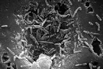
Mycobacterium belong to the family Mycobacterineae of the Actinobacteria. The genus contains pathogens known to cause serious disease in mammals including humans.
Two of the better known diseases are tuberculosis and leprosy.
Lesser known conditions are mycobacterium avium complex (MAC), hypersensitivity pneumonitis, ulcerated skin and muscle lesions, mycetoma, suspected causation of sarcoidosis, among others. Several of these conditions will be discussed below.
A good review of MAC by the American Thoracic Society is published in the American Journal of Respiratory and Critical Care Medicine. Read the paper.
Mycobacterium are aerobic non-motile Gram-positive, being classified as an acid-fast Gram positive straight or slightly curved rods.
All Mycobacterium species have a characteristic thick cell wall, which is hydrophobic and rich in mycolic acids (mycolates).
Many species of the genus adapt very readily to growth on simple substrates, using ammonia or amino acids as a nitrogen source and glycerol as a carbon source. Optimum growth temperature ranges form 25 to over 50 °C. Thus, some are classified as thermophilic and are associated with lung disease.
Mycobacterium can produce pigments and are classified as to the types of pigments as follows:
Group I - Photochromogens: The species produce non-pigmented colonies when grown in the dark and become pigmented after exposures to light and re-incubated. Examples are M. kansasii, M. marinum and M. Simiae.
Group II: Scotochromogens: This group produces deep orange colonies when grown in either light or dark. The species include scrofulaceum, gordonae, xenopi and szulgai.
Groups III & IV: Non-Chromogens: The members of this group are non-pigmented in either dark or light growth conditions. Some may be pale yellow, buff or tan in color. Species of this group include tuberculosis, avium-intra-cellulare, bovis, ulcerans, fortuitum and chelonae .
Mycobacteria are widely spread in the eco-system, being present in water and food sources. They can even persist in chlorinated drinking water. Some species appear to be obligate intracellular parasites, such as in tuberculosis and leprosy.
Several species have been identified in water-damaged building materials and contribute to the indoor air microbial contamination (Andersson et al, 1997; Torvinen et al 2006; Lignell et al, 2005; Rautiala et al, 2004; Pessi et al, 2002).
Pathogenicity and Literature on Diseases Caused by Mycobacterium species
Mycobacteria can colonize without overt symptoms of adverse health effects. A good example of this is M. tuberculosis. Billions of people around the world are infected with this organism but will never know, because they do not develop symptoms characteristic of tuberculosis.
In addition, those individuals who develop the disease may be genetically pre-disposed. Mycobacteria infections are notoriously difficult to treat. Again, tuberculosis, leprosy and infections from M. ulcerans are prime examples.
Because of their thick wall, these bacteria are resistant to antibiotics that attack the integrity of the cell walls, e.g., Penicillin. Also, the cell wall allows resistance to alkalis, acids, detergents, oxidative bursts and other chemical actions that cause disruption of the cell wall and eventual cell death. With this short background, we will now look at some of the health problems associated with Mycobacterium spp.
Mycobacterium Tuberculosis Complex: Several species are causative agents in human and animal tuberculosis. The species are: tuberculosis (human disease), bovis, africanum, canetfi, caprae, micro and pinnipea.
Mycobacterium Avium Complex (MAC): The below introduction is lifted from the American Thoracic Society Review in 2007, mentioned above.
The NTM acronym used below refers to Non Tuberculosis Mycobacterium.
This is the third statement in the last 15 years dedicated entirely to disease caused by WM. The current, unprecedented high level of interest in NTM disease is the result of two major recent trends: the association of NTM infection with AIDS and recognition that NTM lung disease is encountered with increasing frequency in the non-AIDS population.
Furthermore, NTM infections are emerging in previously unrecognized settings with new clinical manifestations.
Another major factor contributing to increased awareness of the importance of NTM as a human pathogen is improvement in methodology in the mycobacteriology laboratory, resulting in enhanced isolation and more rapid and accurate identification of NTM from clinical specimens.
Consistent with the advances in the mycobacteriology laboratory, this statement has an emphasis on individual NTM species and the clinical disease—specific syndromes they produce. A major goal is facilitating the analysis of NTM isolates by the health care provider, including determination of the clinical and prognostic significance of NTM isolates and therapeutic options.
There are controversies in essentially all aspects of this very broad field and, whenever possible, these controversies are highlighted. Hence, an attempt is made to provide enough information so that the clinician understands the recommendations in their appropriate context, especially those made with inadequate or imperfect supporting information.
Also, when there is not compelling evidence for one recommendation, alternative recommendations or options are presented.
When the last ATS statement about NW was prepared in 1997, there were approximately 50 NTM species that had been identified. Currently, more than 125 NTM species have been cataloged.
A list of NM species identified since 1990 is provided in the online supplement.
There has been a dramatic, recent increase not only in the total number of mycobacterial species but also in the number of clinically significant species.
Clinicians might reasonably ask, 'Why are there so many new NTM species?"
The increase relates to improved microbiologic techniques for isolating NTM from clinical specimens and, more importantly, to advances in molecular techniques with the development and acceptance of 165 rRNA gene sequencing as a standard for defining new species.
PATHOGENESIS
Over the past two decades, three important observations have been made regarding the pathogenesis of NTM infections:
1. In patients infected with HIV, disseminated NTM infections typically occurred only after the CD4+ T-lymphocyte number had fallen below 50Ip1, suggesting that specific T-cell products or activities are required for mycobacterial resistance.
2. h the HIV-uninfected patient group, genetic syndromes of disseminated NTM infection have been associated with specific mutations in interferon (IFN)- y and interieulcin (114-12 synthesis and response pathways (FN-y receptor 1 [FNyR1], IFN-y receptor 2 [IFNyR2], IL-12 receptor 131 subunit [11_12R1311, the 1-12 subunit p40 [IL12p40], the signal transducer and activator of transcription 1 ISTAT1], and the nuclear factor-1 essential modulator [NE MC]).
3. There is also an association between bronchiectasis, nodular pulmonary NTM infections and a particular body habitus, predominantly in postmenopausal women (e.g., pectus excavatum, scoliosis, mitral valve prolapse).

