Beta Glucans

Beta Glucans
The 1, 3 beta-D-glucans (glucans) are diagnostic markers for fungal infections, particularly Aspergillus species, and systemic Candidiasis, and other fungi (Kondori et al, 2004; Pickering et al, 2004; Pazos et al, 2005). However, PCR DNA analytical tests give earlier and more specific diagnostic results for infections (Rantakokko-Jalava et al, 2003; Musher et al, 2004; Khan et al, 2007). 

The glucans have been demonstrated in the indoor air and dust and their presence is related to fungal growth and possible intrusion from outdoor sources (Rylander, 1999, 2004; Chew et al, 2001; Gehring et al, 2001; Douwes et al, 2006). The glucans cause airway inflammation. They have been identified in bronchoalveolar lavage fluid from individuals with acute eosinophilic pneumonia (Thorn and Rylander, 1998; Kawayama et aI, 2003). 

Inhalation of glucans by healthy individuals caused an increase in eosinophilic cationic protein, TNF-alpha and a reduction of peripheral blood eosinophil numbers (Thorn et al, 2001). 

Similar observations have been reported in guinea pigs and mice treated with glucans (Fogelmark et al, 2002). In contrast, blood leukocytes from healthy volunteers and patients allergic to house dust glucans enhance the release of IgE and histamine in vitro (Holck et al, 2007). Thus, the effects of inhaled glucans may be different in eosinophilic pneumonia cases vs healthy and allergic individuals when tested in vivo compared to in vitro assays. 

Airway inflammation in adults with chronic exposure to glucans is associated with increased prevalence of atopy, a slight increase in myeloperoxidase and a decrease in forced expiratory volume (FEV1) (Thorn and Rylander, 1998). Moreover, children exposed to glucans in dust at home and at school have variability in pulmonary peak flow values as well as signs of airway inflammation (Rylander et aI, 1998; Rylander, 1997, 1999; Douwes et aI, 2000) and have a higher incidence of infections (Rylander, 2004). 

Finally, nasal deposition of glucans is not associated with acute inflammation with respect to an increase of the chemo-attractant eotaxin and eosinophils in nasal lavage fluid (Beijer and Rylander, 2005). Inflammatory response to 1-3 beta-D-glucans involves TLR2 and TLR4 receptors, MYD88 and Dectin-1 (Wang et al, 2002; Meier et al, 2003; Hohl et al, 2005). 

Finally, antibodies against glucans have been demonstrated in humans and animals exposed to molds (Notermans et al, 1987, 1988; Kamphuis et al, 1992). 

The detection of glucan antibodies suggests that inhalation of glucans and/or colonization/infection has occurred. 

In either event, the antibodies to glucans demonstrate an immune response unrelated to IgE sensitivity. Also, glucans are in the blood of patients with deep invasive mycosis and fungal febrile episodes and can be used to diagnose infections (Miyazaki et aI, 1995; Obayashi et al, 1995; Pickering et aI, 2004; Kedzierska, 2007). 

Additional work is needed to determine the role of glucans in respiratory inflammation. However, children seem to be more susceptible to exposure. 
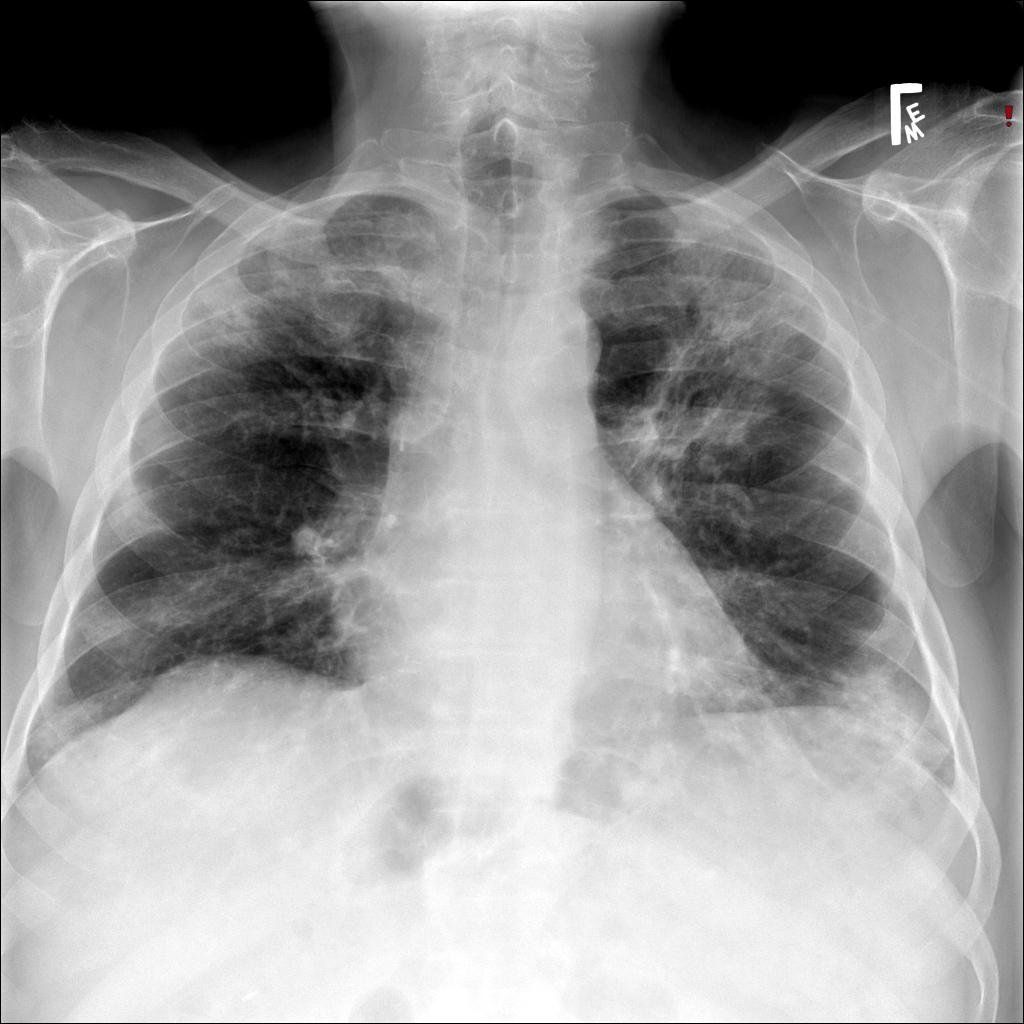
Lung x-ray showing acute eosinophilic pneumonia

Lung x-ray showing acute eosinophilic pneumonia


Share by: