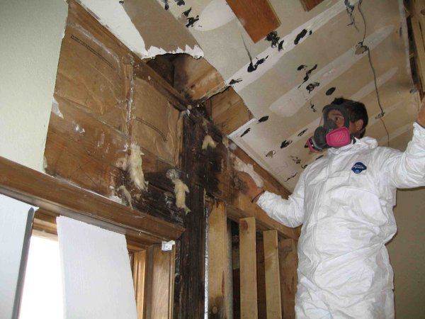
Fungal contamination as a major contributor to sick building syndrome has been reviewed. The significant factors for mold growth are water, temperature and substrate (Li and Yang, 2004). Water activity (aw) represents available water in a substrate. It is expressed as a decimal fraction of the amount of water present in a substrate that is in equilibrium with relative humidity.
Molds that grow at various aw are classified as xerophilic (xerotolerant), mesophilic and hydrophilic. The xerophilic molds include species of Penicillium, Aspergillus and Eurotium that grow at aw <0.8. Mesophilic molds grow at aw 0.8-0.9 and include Alternaria, Cladosporium, Phoma, Ulocladium and Epicoccum nigrum. The hydrophilic molds include Chaetomium globosum, Fusarium, S. chartarum, Memnoniella echinata, Rhizopus stolonifer and Trichoderma spp. at aw >0.9.
Thus, the genera of mold identified indoors are indicative of the extent of water intrusion. Ergosterol and mycotoxins are indicators of mold growth (Hippelein and Rugamer, 1984; Li and Yang, 2004).
Molds grow on surfaces as well as in hidden areas such as in carpet, behind wall paper, inside interior and exterior walls, in attics, in subflooring, etc. They thrive on wet building materials rich in carbohydrates.
Molds take nutrients from dead organic material (wood, dry wall, paint, paper, glues, etc.) by secreting digestive enzymes into the matrix upon which they are growing. It is estimated that approximately 50% of the mold growth in damp indoor environments is hidden, e.g., within wall cavities, carpets, etc.
Molds are present in the ventilation systems of contaminated homes, buildings and automobiles (Li and Yang, 2004; Ahearn et al, 1996, 2004).
Certain species of molds are more abundant (amplified) indoors vs outdoors. These include Aspergillus flavus, versicolor, sydowii, niger and fumigatus and Penicillium chrysogenum, brevicompactum, citrinum and decumbens, Chaetomium, Epicoccum, Fusarium and S. chartarum. Cladosporium species are often equally abundant outdoors and indoors.
The comparison of total mold spore counts from indoor to outdoor samples is not an adequate method to test for mold contamination. Air sampling is only a snapshot of an indoor environment that fluctuates according to various parameters, e.g., human activity, air conditioning, temperature, opened/closed windows, etc. The extent of mold and other microbial growth must be determined from a combination of samples that include bulk, wipe, air, carpet dust and wall cavity
sampling.
Next, the frequency (percentage) of various species of Aspergillus, Penicillium, Stachybotrys, etc., in the indoor vs the outdoor samples must be determined. This approach will reveal that several of the aforementioned molds have amplified indoors when compared to outdoors.
The profile of indoor mold may be constant when compared to the outdoor profile. However, certain species of Aspergillus and Penicillium, as mentioned above, will be dominant indoors vs outdoors (Schwab and Straus, 2004; Straus et al, 2003; Wilson and Straus, 2002). For example, the authors have observed situations in which Penicillium species were at 100% in the indoor air and bulk samples, while outdoor levels were <12%. Moreover, in other samples, Aspergillus species were greater indoors (46%) vs outdoors (<6%).
The US EPA has developed the polymerized chain reaction (PCR) DNA technology to identify 130 of the major indoor fungi to the species level and has licensed several companies to utilize the method (USEPA, 2007).
As an example, the role of air speed equivalent to normal human activity on the release of spores from mold colonies has been reported in bench studies (Gorny et al, 2001, 2002; Gorny 2004; Tucker et al, 2007). Low air speeds cause an initial release of spores from colonies of S. chartarum, Aspergillus niger and versicolor; Penicillium chrysogenum and melinii, and Cladosporium sphaerospermum and cladosporioides. The spore release per square centimeter during the first ten minutes was lowest for C. cladosporioides (<500 spores); followed by S. chartarum (4,000 spores) and Aspergillus and Penicillium spp. (10,000 spores each).
After the initial release, additional spore releases did not occur with Stachybotrys and Cladosporium, while those of Aspergillus and Penicillium slowly declined for the duration of the 70 minutes of observations (Tucker, et al, 2007). The proportion of released amounted to 0.2% (S. chartarum); 0.8% (C. cladosporioides); 1.1% (A. niger) and 1.8% (P. chrysogenum) of total spore mass of each mold. These observations demonstrate that dry spore molds (Aspergillus and Penicillium) more readily release their conidia when compared to sticky clusters of spores of S. chartarum.
Moderately to heavily damaged homes in the aftermath of Katrina had elevated levels of Aspergillus, Penicillium and Paecilomyces (Chew et al, 2006; Rao et al, 2007b). Moreover, molds detected in water-damaged building materials include A. versicolor, A. sydowii, Trichoderma viride, S. chartarum, Chaetomium globosum , multiple species of Penicillium, Acremonium, Cladosporium, Phoma, Aureobasidium, Phialaphora and yeast (Reijula, 2004).
Rodent lungs have been used to test the adverse effects of various components of common indoor molds. Proteases, isosatratoxin-F, and spores of S. chartarum cause inflammatory and cytotoxic effects in the lungs of juvenile mice. Among the adverse effects are alterations and morphological changes in Type II alveolar cells (Rand, et al, 2002), release of IL-1-beta, IL-6, IL-8 and TNF-alpha with neutrophilia, granuloma and reduced collagen IV (Yike et aI, 2007; Pestka et al, 2008) and a decrease in alveolar space (Rand, et aI, 2003).
Moreover, spores from indoor species of Aspergillus and Penicillium cause lung eosinophilia, neutrophilia, release of inflammatory cytokines (IL-6, TNF-alpha), vascular leakage, elevated LDH, Th-2 inflammatory responses and other cytotoxic damage in mouse lungs (Jussila et al, 2002b; Schwab and Straus, 2004; Cooley et aI, 1998, 2000, 2004).
Stachybotrys chartarum consists of two chemotypes: one produces trichothecene mycotoxins, while the other releases spirocyclic atranones. Both cause inflammation in mouse lungs (Flemming et al, 2004). Stachybotrys chartarum is a slimy greenish black mold that does not readily release spores. Thus, the presence of its spores in the indoor air and bulk samples signals either dried disturbed colonies and/or contamination. The spores of Stachybotrys are rare findings in outdoor air (Cooley et al, 1998; Shelton et al, 2002; Li and Yang, 2004; Schwab and Straus, 2004).
Another issue that has been overlooked is the role of molds in chronic upper and lower respiratory tract disease. Chronic rhinosinusitis (CRS) appears to be a non-lgE immunological inflammatory response to fungi with nasal eosinophilia and the release of toxic major basic protein and has a favorable response to intranasal amphotericin B (Sasama et al, 2005; Ponikau et al, 2005, 2006; Kern et al, 2007). Cladosporium, Aspergillus, Alternaria, and Penicillium were frequently cultured from nasal polyps with a histologic type of fibro inflammation present in over 60% of patients vs controls. Fungi were commonly cultured during the hot and humid environment of summer time from the polyps (Shin et al, 2007).
In addition, Alternaria, Aspergillus, and Cladosporium proteases interact with nasal epithelial cells activating protease receptors (PARs 2 and PARs 3) enhancing the production of chemical mediators and migration of eosinophils and neutrophils into nasal polyps (Shin et al, 2006) The condition involves exaggerated humoral response of both TH1 and TH2 types. Peripheral mononuclear blood cells (PMBCS) from CRS patients produce significantly elevated IL-5 and IL-13 to Alternaria and Cladosporium antigens and increased levels of IgG antibodies to Alternaria (Shin et al, 2004). Also, the antimicrobial peptide, cathelicidin LL-37, is up-regulated by antigens of Aspergillus (4 fold) and Alternaria (6 fold) in CRS patients as well as up-regulated surfactant protein (Ooi, et al, 2007a,b).
Molds may infect and/or colonize. Rao et al, (2007a) reported 8 cases of colonization of New Orleans immunocompetent residents with Syncephalastrum. The organism was isolated from clinical specimens of sputum, BAL, endotracheal aspirates and nasal swabs. Also, the existence of aspergillosis in immunocompetent and immune compromised humans is not questioned (Strelling et al, 1966; Lewis et al, 2005 a, b; Raja and Singh, 2006; Samarakoon and Soubani, 2008).
Several other Zygomycetes are capable of causing human disease (Ribes et al, 2000). Mucormycosis of immunocompetent individuals with involvement of the gastrointestinal tract, skin, paranasal sinuses, necrotizing fasciitis and pericardium has been described in India (Jain et al, 2006; Prasad et al, 2008).
Pulmonary aspergillosis and clinical update of the disease has been recently reviewed (Zmeili and Soubani (2007). The recognized clinical conditions are aspergilloma, pulmonary aspergillosis (noninvasive), invasive aspergillosis (IA), chronic necrotizing aspergillosis (CNA) and allergic bronchopulmonary aspergillosis (APBA). 00 (fungus ball) occurs in individuals with pre-existing lung disease (tuberculosis, sarcoidosis, bronchiectasis and cysts).
The fungus ball exists in pre-existing cavities in diseased lungs. It contains fungal hyphae, inflammatory cells, fibrin, mucous and tissue debris. Other organisms causing fungal balls are Zygomycetes and Fusarium. Diagnosis of pulmonary aspergilloma is usually based upon clinical history and radiographic features.
Approximately 50% of sputum cultures are positive for fungi. The development of IA is usually rapid in immunocompromised individuals (e.g., cancer chemotherapy, organ transplant and hematopoietic stem-cell transplantation) with high mortality in neutropenic (50%) and HSCT (90%) patients. CNA is semi-invasive resulting from infections of the lungs and runs a slow progressive course.
APBA is a hypersensitivity to Aspergillus antigens usually in individuals with asthma or cystic fibrosis who are dependent upon chronic corticosteroid therapy. APBA is an inflammatory condition involving Hypersensitivity Type I and III immune responses. The pathology is poorly understood while chronic granulomatous conditions exist in the lungs of affected individuals.
IA by Aspergillus fumigatus have been reported in immuno-competent children. Strelling et al (1966) describe the deaths of a brother (14 months) and sister (4 yrs) from acute pulmonary aspergillosis and review the literature. The two children developed acute fever, cough and breathlessness with yellowish-brown (hemoptysis) mucus after playing in a barn. Cultures from the lungs and barn materials isolated A. fumigatus, while tissue sections of the lungs revealed branching fungal hyphae. The doctors’ review of the literature described additional deaths and disease from aspergillosis as follows: 1) brother (5 yrs) and sister (11 yrs) living on a farm; 2) 20-day-old infant; 3) 18-day-old infant; 4) 7-yr-old child (sex not given); 5) Boy (6 yrs); and 6) girl (7 yrs) who recovered. The cerebellum and frontal lobes were involved in cases 5 and 6.
Immunocompetent adults may develop noninvasive or invasive Aspergillus infections. Noninvasive aspergillosis involves aspergilloma of the lungs visible on CT scan with a solitary nodule or mass. Microscopically granuloma and cavity lumen hyphae are present (Kang et al, 2002). On the other hand, invasive aspergillosis (IA) may involve the lungs as well as infecting other organs. The lungs can be almost completely consolidated with granuloma (Hillerdal et al, 1984; Reijula and Tuomi, 2003), bilateral hilar prominence (Parameswaran et al, 1999), bilateral fibrinous pleural adhesion and extensive parenchymal destruction (Zuk et al, 1989).
Blood stained mucous in bronchi and massive haemoptysis may be also present (Parameswaran et al, 1999; Zuk et al, 1989).
Invasion of the tracheobronchial tree can also occur (Mohan et al, 2005). Individuals with asthma or COPD on either short-term or prolonged corticosteroid therapy (oral, inhalation, I.V.) contract IA mostly from A. fumigatus followed in order by flavus, terreus, niger and nidulans (Ganassini and Cazzadori, 1995; Smeenk et al, 1997; Ali, et al, 2003; Trof et aI, 2007; Samarakoon and Soubani, 2008).
Disseminated IA to the skin and bone (spondylodiscitis) in a patient with a pulmonary nodule has been described (Domergue et al, 2008), while another documented patient involved the lung, liver and spleen (Raja and Singh, 2006). Paranasal sinus involvement, perforation of the nasal septum, and invasion of the palate was described in one patient (Khatri et al, 2000; Raja and Singh, 2006; Samarakoon and Soubani, 2008), while another had paranasal, orbital involvement with intracranial extradural extension via the maxillary division of the trigeminal nerve (Subramanian et al, 2007). IA can result in dissemination to the central nervous system (CNS) (Garcia et al, 2006; Palanisamy et al, 2005).
CNS complications in one person involved headache, nausea, motor impairment and cognitive decline due to progressive cerebellar lesions. After treatment with high doses of itraconazole (1600 mg/day), the patient recovered with only mild cerebellar motor impairment (Palanisamy et al, 2005).

