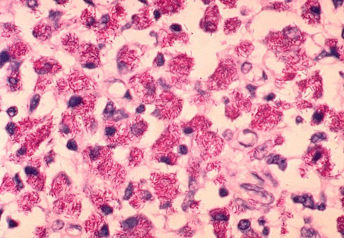
Gram positive bacteria have been isolated from water-damaged
building materials. Actinomycetes (Actinobacteria), including several species of Streptomyces, Nocardia and Mycobacterium, were cultured from indoor air and dust as well as from moldy, water-damaged materials (Peltola et al, 2001a, b; Rintala et al, 2001, 2002, 2004; Rautiala et al, 2004; Torvinen et al, 2006).
Several species of both potentially pathogenic and saprophytic Mycobacteria were isolated from the workplace air during remediation (Rautiala et al, 2004). Some of the identified species belonged to the Mycobacterium avium complex and are potential human pathogens. It was noted by the authors that the mycobacteria are slow growing.
The Actinomycetes are potential human pathogens. The reporting of these infections in the U.S. is not required; therefore, it is impossible to determine disease prevalence.
Streptomyces californicus produces spores approximately 1 micron in mean aerodynamic diameter that can penetrate deep into alveolar spaces of the lungs. Intrathecal instillation of spores in mice caused inflammation characterized by increase concentrations of TNF alpha, IL6, LDH, albumin, and hemoglobin in bronchoalveolar lavage fluid (BAL) and sera (Jussila et al, 2001).
Moreover, following repeated exposures to the spores in a dose response study, the inflammation was systemic--involving recruitment of neutrophils, macrophages and activated lymphocytes into the airways and decreased numbers of spleen cells. The dose response was nonlinear.
BAL from the mice also contained increased concentrations of albumin, total protein, LDH and activated lymphocytes. It was concluded that S. californicus spores are capable of causing both lung inflammation and systemic immunotoxic effects (Jussila et al, 2002a, 2003).
It is interesting that Streptomycetes produce anthracyclines, (e.g., daunorubicin and doxorubicin) drugs widely used in chemotherapy (Arcamone, 1998; Arcamone and Cassinelli, 1998). The anthracyclines cause apoptosis of activated and nonactivated lymphocytes as well as a decrease of mature T and B cells in mice. The T and B cell depletion was most severe in the spleen, moderate in lymph nodes and least in the thymus (Ferraro et al, 2000).
In addition, various species of this genus are the source for a variety of antibiotics (Gunsalus, 1986).
The inflammatory and cytotoxic affects in vitro of indoor air bacteria compared to mold spores has been reported. The bacteria tested were Bacillus cereus, Pseudomonas fluorescens, and the molds tested were Streptomyces californicus, A. versicolor, P. spinulosum, and S. chartarum. The bacteria caused the production of IL6 and TNFalpha in mouse macrophages.
Only the spores of S. californicus caused a production of nitric oxide (NO) and IL6 in both mouse and human cells. Of the molds, only S. chartarum caused the production IL6 in human cells. The overall potency to stimulate the production of proinflammatory mediators decreased in order as follows: Ps. fluorescens, S. californicus, B. cereus, S. chartarum, A. versicolor, P. spinulosum. There was a synergistic response of TNFalpha and IL6 after coexposure with S. californicus with both trichodermin and 7alphahydroxytrichodermol.
These observations indicate that bacteria in water-damaged
buildings should also be considered as causing inflammatory effects on occupants (Huttunen et al, 2004).
The synergism and interaction of S. californicus and S. chartarum on mouse macrophages have been reported. Spores from these two organisms were tested for the effects on macrophages as follows: spores isolated from co-cultivated cultures, mixture of the spores from separate pure cultures and the spores of each organism. Spores isolated from the co-cultures were compared to the mixture of spores and were more cytotoxic than either the mixture of spores or the spores from each organism. Co-cultured spores caused increased apoptosis of the macrophages by more than 4fold.
Cells arrested at G2/M stage of the cell cycle were increased nearly two-fold. In contrast, the co-cultured spores significantly decreased the ability of the spores to trigger the production of NO and IL-6 by the macrophages. Thus, co-culturing of the two organisms resulted in microbial interactions that significantly potentiated the ability of spores to cause apoptosis and cell cycle arrest (Penttinen, et al, 2005).
In a follow-up study, the same authors (Penttinen et al, 2006) compared the cytotoxicity of the co-cultured spores (S. californicus and S. chartarum) to that of chemotherapeutics (doxorubicin, phleomycin, actinomycin D and mitomycin C) produced by S. californicus. The co-cultured spores mediated apoptosis, cell cycle arrest at the S-G2/M phase and caused a 4-fold collapse of mitochondrial membrane potential.
In addition, a 6-fold increase in caspase-3 activation and DNA fragmentation was observed. The cytotoxicity of the co-cultured spores was similar to that caused by doxorubicin and actinomycin D. It was concluded that the co-culture of the two organisms caused the production of unknown cytotoxic compound(s) which evoked immunotoxic effects similar to chemotherapeutic drugs. In conclusion, these studies demonstrate that spores from S. californicus are cytotoxic to mouse macrophages.
More importantly, the co-cultivation of S. californicus and S. chartarum results in a spore mixture that is more toxic than the spores of each organism cultured individually.
Additional attention must be paid to the synergism that probably occurs in the microbial mixture that is present in water-damaged buildings.
Moreover, the spores of S. californicus were more toxic to mouse macrophages than was a mixture of spores from co-cultures with various molds (A. versicolor, P. spinulosum and S. chartarum). S. californicus spores alone were more potent inducers of inflammatory and cytotoxic responses than any combination of co-cultivated spore mixtures.
In addition, co-culture of S. chartarum and A. versicolor produced a synergistic increase in cytotoxicity with no effect on inflammatory responses of the macrophages (Murtoniemi et al, 2005).
Finally, S. griseus strains isolated from indoor environments produce a toxin, valinomycin, which causes mitochondrial swelling, damaged mitochondrial membranes and disrupted the mitochondrial membrane potential of boar sperm (Andersson, et al, 1997; Peltola et aI, 2001a).
The Nocardiopsis strains isolated from indoor water-damaged environments are toxigenic and produce a mitochondrial toxin that damages the mitochondria of boar sperm (Peltola et al, 2001a, b). In conclusion, these studies demonstrate that spores from S. californicus are cytotoxic to mouse macrophages.
More importantly, the co-cultivation of S. californicus and S. chartarum results in a spore mixture that is more toxic than the spores of either organism cultured individually. Additional attention must be paid to the synergism that probably occurs in the microbial mixture that is present in water-damaged buildings.
Finally, Nocardia isolated from water-damaged building materials also cause cytotoxicity.
Streptomyces species are associated with farmer's lung disease (allergic alveolitis). Infections (Streptomycosis) occur most frequently in immunocompromised individuals and people with diabetes mellitus and/or corticosteroid therapy. However, co-infection with Aspergillus in cases of chronic granulomatous disease following exposure to aerosolized mulch has been reported (Siddiqui et al, 2007). Diagnosis is difficult because mimicry of other diseases (Kagen et al,1981; Che et al,1989; Roussel et al, 2005; Kapadia et aI, 2007; Kofteridis, et al, 2007; Madhusudhan et aI, 2007; Acevedo et aI, 2008; Quintana et aI, 2008).
Finally, Streptomyces spp. can cause mycetoma, a condition most endemic around the Tropic of Cancer, but also occurs worldwide, in the U.S., Asia, and Latin America (Welsh et al, 2007; Quintana et al, 2008). The organisms are aerobic, producing chalky aerial mycelia and produce granules of different sizes, textures and colors. Streptomyces do not stain with hematoxylin and eosin, but are gram positive and acid fast stain.

