Sarcoidosis

Sarcoidosis
Sarcoidosis is a disease that can involve all organs of the body. The causes of the disease are slowly being revealed in the current scientific and medical literature. 

Sarcoidosis is a granulomatous inflammatory disease that occurs in lymph nodes, lungs, liver, eyes, skin, joints, brain and other organs. The inflammation results in granulomas in these organs that are driven by TH1/TH17 lymphocytes, dendritic cells, chemokines and pro-inflammatory cytokines. 

Who has Sarcoidosis? 

Once thought rare, sarcoidosis is now known to be common and affects people worldwide. The disease can affect people of any age, race and gender. However, it is most common among adults between the ages of 20 and 40 and in certain ethnic groups.
  • In the United States, it is most common in African Americans and people of European "particularly Scandinavian" descent. 
  • Among African Americans, the most affected U.S. group, the estimated lifetime risk of developing sarcoidosis might be as high as 2 percent. 
  • Most studies suggest a higher disease rate for women. 
Disease severity can vary by race or ethnicity.
  • Higher among African Americans than among Caucasians.
  • Appearance of the disease outside of the lung is common in certain populations. For example: the eyes (chronic uveitis) in African Americans, painful skin lumps in Northern Europeans, and the heart and eyes in Japanese.
What are the Symptoms? 

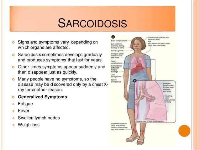
Sarcoidosis is a multi-system disorder. Symptoms typically depend on which organ the disease affects. Most often, the disease will affect the lungs. The symptoms and organs involved in sarcoidosis can vary from individual to individual.
  • General: About one third of patients will experience non-specific symptoms of fever, fatigue, weight loss, night sweats and an overall feeling of malaise (or ill health).
  • Lungs: The lungs are affected in more than 90% of patients with sarcoidosis. A cough that does not go away, shortness of breath, particularly with exertion and chest pain occur most frequently with the pulmonary form of the disease. 
  • Lymph Nodes: Up to 90% of sarcoidosis patients have enlarged lymph nodes. Most often they are in the neck, but those under the chin, in the arm pits and in the groin can be affected. The spleen, which is part of the lymphatic system, can also be affected. 
  • Liver: Although between 50% to 80% of patients with sarcoidosis will have granulomas in their liver, most are without symptoms and do not require treatment. 
  • Heart: Researchers estimate that cardiac sarcoidosis, affects more than 10 percent of people with sarcoidosis in the United States, and perhaps as many as 25 percent. Sarcoidosis can cause the heart to beat weakly resulting in shortness of breath and swelling in the legs. It can also cause palpitations (irregular heartbeat).  
  • Brain & Nervous System: From 5% to 13% of patients have neurologic disease. Symptoms can include headaches, visual problems, weakness or numbness of an arm or leg and facial palsy. 
  • Skin: One in four (25%) patients will have skin involvement. Painful or red, raised bumps on the legs or arms (called erythema nodosum), discoloration of the nose, cheeks, lips and ears (called lupus pernio) or small brownish and painless skin patches are symptoms of the cutaneous form of the disease.
  • Bones, Joints & Muscles: Joint pain occurs in about one-third of patients. Other symptoms include a mass in the muscle, muscle weakness and arthritis in the joints of the ankles, knees, elbows, wrists, hands and feet. 
  • Eyes: Any part of the eye can be affected by sarcoidosis and about 25% of patients have ocular involvement. Common symptoms include: burning, itching, tearing, pain, red eye, sensitivity to light (photophobia), dryness, seeing black spots (called floaters) and blurred vision. Chronic uveitis (inflammation of the membranes or uvea of the eye) can lead to glaucoma, cataracts and blindness. 
  • Sinuses, Nasal Muscosa (lining) & Larynx: About 5% of patients will have involvement in the sinuses with symptoms that can include sinusitis, hoarseness or shortness of breath. 
  • Other Organs: Rarely, the gastrointestinal tract, reproductive organs, salivary glands and the kidneys are affected. 
Diagnosis

Physical examination may reveal the following: enlarged liver and spleen, enlarged lymph nodes (neck and axillary), skin rashes or sores.

Symptoms of sarcoidosis can mimic those of other diseases. In some cases, sarcoidosis may be diagnosed by excluding these other similar diseases. Frequently, sarcoidosis is diagnosed because a routine chest x-ray shows an abnormality. To accurately diagnose the disease, most doctors will take a medical history and perform a physical examination. Laboratory tests of the blood, chest x-rays, breathing tests and biopsy are all diagnostic tools.

The disease can be progressive (e.g., beginning with stage I through IV). Stage IV is almost always systemic.

References

1. Berge BT, Kleinjan A, Muskens F, Hammad H, et al. 2012. Evidence for local dendritic cell activation in pulmonary sarcoidosis. Respir Res 13:33.

2. D’Angel C, De Luca A, Zelante T, Bonifazi P et al. 2009. Exogenous pentraxin 3 restores antifungal resistance and restrains inflammation in murine chronic granulomatous disease. J Immunol 183:4609-18.

3. Facco M, Basesso L, Miorin M, Bortoli M, et al. 2007. Expression of CCR/CCL20 chemokine axis in pulmonary sarcoidosis. J Leukocyte Biol 82:946-55.

4. Gupta D, Agarwal R, Agarwal AN, Verma I. 2011. Immune responses to mycobacterial antigens in sarcoidosis: A systematic review. 53:41-9.

5. Tercelj m, Stopinsek S, Ihan A, Salobir B et al. 2011. In vitro and in vivo reactivity to fungal cell wall agents in sarcoidosis. Clin Exper Immunol 166:87-93.

6. Zelante T, Bozza S, De Luca A, D’Angelo C et al. 2009. Th 17 in the setting of Aspergillus infection and pathology. 47 (Suppl 1) S162-9.

7. Zelante T, De Luca A, Bonifazi P, Montagnoli C, et al. 2007. Il-23 and the Th 17 pathway promote inflammation and impair antifungal immune resistance. Eyr J Immunol 37:2695-706.
8. Sarcoidosis defined by NIH. Read the article.  

9. Histology of Granulomas. Go to the website
Global Indoor Health Network - Sarcoidosis caused by environmental exposures

Sarcoidosis and environmental exposures


Sarcoidosis and Mold

Sarcoidosis has been associated with several different work and environmental exposures such as agricultural employment, insecticides at work and work environments with mold and mildew as reported by Newman, et al. 

A separate investigation of respiratory morbidity in office workers in a a water-damaged building also reported a correlation between respiratory disease (asthma, Hypersensitivity Pneumonitis) and Sarcoidosis. The asthma rate increased from 1.9/1000 person years before occupancy to 14.5/1000 persons years after occupancy. Cases of HP and sarcoidosis were also associated with occupancy. 

The question that arises is: What organisms are associated with sarcoidosis? 

Recently, Aspergillus flavus and fumigatus were identified in the lungs of 10 patients from a retrospective cohort of 447 cases of sarcoidosis. The limitation of this report was that environmental exposures that may have led to the sarcoidosis in all patients was not done. Thus, it cannot be said which came first-- Aspergillus or sarcoidosis--as casually related to the disease. However, other published data show a causal relationship between pulmonary granulomas (sarcoidosis) and fungal infections.

Mukhopadhyay, et al, published their findings regarding causative agents in pulmonary granulomas in 500 patients with pulmonary granulomas. Specific cause was identified in 58 % of the cases (sarcoidosis in 27% and mycobacterial and fungal 28% of the 290 cases). Insufficient information was available for identification of cause in 210 of the 500. 

Fungi were identified in biopsy specimens, while Mycobacteria were identified by culture. The identified mycobacterium will be discussed in the review of bacteria infections in sarcoidosis published on this web site. Of interest, Mycobacteria were identified in 19% of the cases outside of the USA versus 8% within the USA. 

In contrast, fungi accounted for 19% of cases in the USA versus 4% in other countries. No attempt was made to identify possible environmental sources of the microbial infections, e. g., workplace, water-damaged buildings. 

Nonetheless, fungal and bacterial infections were identified as the probable cause of granulomas in these cases. However, several more recent papers have appeared in the literature that indicate that fungal exposure is causally related to the development of sarcoidosis. These data will be briefly reviewed below.

Fungi and Sarcoidosis

Increasing evidence has shown a positive correlation between exposure to microbes (particularly molds and Mycobacterium) and the hypersensitivity reaction leading to the development of granulomas and sarcoidosis. 

Sarcoidosis historically has been treated with corticosteroids. However, some patients are refractory to this treatment. 

Initially, a pilot study of refractory subjects involving 18 patients with sarcoidosis grades I and II was treated with antifungals (itraconazole, fluconazole, and ketonazole (all at 200 mg/day) along with corticosteroids. At 3-6 months, treatment resulted in a significant decrease of pulmonary infiltration. In addition the patients had significant increases in diffusion capacity and decreases in the severity of symptoms. The study suggested that antifungals are useful in certain cases of sarcoidosis associated with Aspergillus and Candida circulating antibodies. 

A more recent study involved newly diagnosed sarcoidosis (n = 92) patients as follows: corticosteroids (n = 39), corticosteroids + antifungals (n = 31) and antifungals (n = 22). The patients had circulating antibodies against Aspergillus and Candida as well as biomarkers indicative of sarcoidosis (Angiotensin-converting enzyme and chitotriodases. X-ray score decreased significantly among all three groups, while antifungal treatment groups had greater improvement in X-ray findings when compared to the corticosteroid groups (p ,<0.001). It was concluded that antifungal treatment was as efficient as corticosteroid treatment against granulomatous and inflammatory manifestations of sarcoidosis.

Two additional observations support the role of fungi in the development of sarcoidosis. N-acetylhexosaminidase (NAHA), a fungal cell wall enzyme is a biomarker of fungal biomass and indoor mold exposure. NAHA levels were tested in homes where individuals had active and/or recurrent sarcoidosis. 

Individuals with active and/or recurrent sarcoidosis had higher activities of NAHA in their homes vs control homes. The values were 33.6-39.9 U/cubic meter (patients) vs 10 U/cubic meter (controls). The second report involved the testing for proinflammatory cytokines in patients with sarcoidosis as well as the proinflammatory response of peripheral blood monocytes stimulated by particulate and soluble (S-glucan and P-glucan) Beta-glucans, chitin and lipopolysaccharide (LPS). The cytokines tested were TNF-alpha, IL-2, IL-10 and IL-12 in vivo and in vitro. The study involved 22 sarcoidosis patients vs 20 controls. 

Briefly, the induced secretion of cytokines was higher from the peripheral blood mononuclear cells from subjects with sarcoidosis. P-glucan was more potent than soluble glucan (S-glucan) for inducing cytokine release. Among the subjects with sarcoidosis, there was a significant relation between the spontaneous production of LI-6, IL-13 and the NAHA levels in the homes. The X-ray findings of the Sarcoidosis patients were related to an increased secretion of cytokines after stimulation with LPS (endotoxins) and P-glucan. Subjects with sarcoidosis had a higher reactivity to the fungal cell wall agents in vitro and to home exposure. 

Closing Comments: Although the above observations clearly implicate particulate glucans in the development of sarcoidosis, other factors must also be considered. These are (1) fine particulates less than one micron releases that contain beta-glucans and secondary metabolites of both mold and bacteria (12-16); and 2) multiple virulence factors have a role in the pathogenesis of both mold and bacteria in the development of lung disease and infection. 

References

1. Newman LS, Rose CS, Bresnitz EA, Rossman MD, et al. 2004. A case control etiologic study of sarcoidosis. Am J Respir Crit Care Med 170:1324-30.

2. Cox-Ganser JM, White SK, Jones R, Hilsbos K, et al. 2005. Respiratory morbidity in office workers in a water-damaged building. Environ Health Perspec 113:485-90.

3. Pena TA, Soubani AO, Sumavati L. 2011. Aspergillus lung disease in patients with sarcoidosis: A case series and review of the literature. Lung 189:167-72.

4. Mukhopadhyay S, Farver CF, Vaszar T, Dempsey OJ, et al. 2012. Causes of pulmonary granulomas: a retrospective study of 500 cases from seven countries. J Clin Pathol 65:51-57.

5. Marshall TG, Marshal FE. 2004. Sarcoidosis succumbs to antibiotics – implications for autoimmune disease. Autoimmunity Reviews 3:295-300.

6. Tercelj M, Salobir B, Rylander R. 2008. Microbial antigen treatment in sarcoidosis- a new paradigm? Medical Hypothesis; 70:831-4.

7. Tercelj M, Rott T, Rylander R. 2007. Antifungal treatment in sarcoidosis – a pilot study. Respir Medicine 10:74-8.

8. Tercelj M, Salobir B, Aupancic K. Rylander R. 2011. Antifungal medication is efficient in the treatment of sarcoidosis. Ther Adv Respir Dis. 2011 Jun;5(3):157-62. Epub 2011 Mar 24.

9. Rylander R, Reeslev, Hulander T. 2010. Airborne enzyme measurements to detect indoor mould exposure. J Environ Monit 12:2161-4. 

10. Tercelj M, Salobir B, Harlander M, Rylander R. 2011. Fungal exposure in homes of patients with sarcoidosis – an environmental study. Environ Health 20:8.

11. Tercelj M, Stopinsek S, Ihan A, Salobir B. et al. 2011. In vitro and in vivo reactivity of fungal cell wall agents. Clin Exper Immunol 166:87-93.

12 Gorny RL. 2004. Filamentous microorganisms and their fragments in indoor air. Ann Agric Environ Med 11:185-97.

13. Reponen T, Seo S-C, Grimsley F, Lee T, et al. 2007. Fungal fragments in moldy houses houses: A field study in homes in New Orleans and Southern Ohio. Atmos Environ 41:8240-9.

14. Brasel RL, Douglas DR, Wilson SC, Straus DC. 2005. Detection of airborne Stachybotrys chartarum Macrocyclic Trichothecenes Mycotoxins on particulates smaller than conidia. Appl Environ Microbiol 71:114-12.

15. Straus DC. 2011. The possible role of fungal contamination in sick building syndrome. Frontiers Bioscience E3:562-80.

16. Taubel M, Sylyok M, Vishwanath V, Bloom E, et al. 2011. Co-occurrence of toxic bacterial and fungal secondary metabolites in moisture-damaged environments. Indoor Air; Oct 21(5):368-75. Epub 2011 May 18.

17. Abad A, Fernandez-Molina JM, Bikandi J, et al. 2010. What makes Aspergillus fumigatus a successful pathogen? Genes and molecules involved in invasive aspergillosis. Revista Iberoam Micol 27:135-82. 

18. Mitchell CG, Slight J, Donaldson K. 1997. Diffusible component of Aspergillus fumigatus which inhibits macrophage oxidative burst is distinct from gliotoxin and other hyphal toxins. Thorax 52:796-801.

19. Griffith DE, Aksamit T, Brown-Elliot BA, Catanzaro A, et al. 2007. An Official ATS/IDSA Statement: Diagnosis, treatment, and prevention of nontuberculous mycobacterial infections. Am J Respir Crit Care Med 175:367-416.
Share by: